
With the launch of EmboGuide during APCCVIR and CIRSE in 2014, Philips introduced a clinical solution for Interventional Oncology that aims to help clinicians more effectively target tumors and their feeding vessels during trans-arterial chemo-embolization (TACE) procedures. * EmboGuide is 510(k) pending, not available for sale in the USA
EmboGuide – at the heart of OncoSuite
EmboGuide is an interventional tool of OncoSuite, the world’s first complete solution that supports embolization and ablation oncology procedures in one room. OncoSuite features the AlluraClarity FD20 and Allura Xper FD20 interventional X-ray systems with a new 3D imaging chain. Its unique technologies and tools support clinicians to better see, reach, and assess tumors.
EmboGuide – workflow-based embolization guidance
Detecting small (less than 2 centimeters) hepatocellular carcinomas (HCCs) during TACE procedures is difficult using conventional 2D digital subtraction angiography (DSA) because of limited tumor vascularity or overlapping vessels.¹ ² Leveraging the fast, high quality 3D imaging of XperCT Dual, EmboGuide provides the first workflow-based tool to guide the detection and treatment of tumors and vessel feeders to multiple lesions in three easy steps.
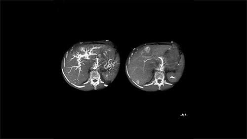
Step 1 – See
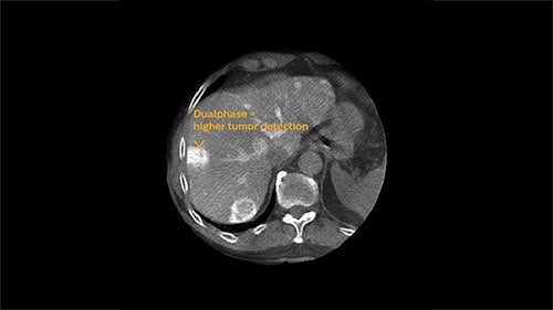
XperCT Dual allows two phases of a contrast injection to be displayed next to each other. Clinicians can assess the feeder vessels to a tumor in the arterial phase and see the tumor boundaries in the delayed phase. Studies have shown that this novel method is superior to conventional DSA and provides equivalent imaging information to the gold standard of contrast enhanced MRI.³ ⁶
EmboGuide is based on XperCT Dual, a dedicated type of 3D oncologic imaging, which is used to visualize tumors and their feeding vessels.
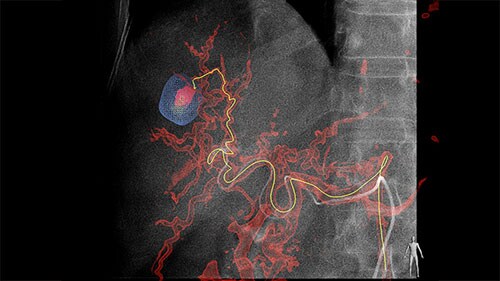
Step 2 – Reach
Based on these images, EmboGuide’s automatic feeder detection can detect significantly more feeding vessels than the current treatment standard of DSA.² ⁴
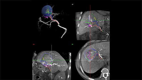
Live Image Guidance then supports selective or super-selective approaches to the embolization target.
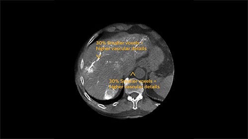
Step 3 – Assess
5 sec. Fast acquisition = reduces breathing artifacts
At the end of each procedure the dedicated oncologic imaging of XperCT Dual helps to determine the treatment endpoint in TACE for HCC.⁵ With XperCT Dual’s fast acquisition protocol, high resolution and soft-tissue contrast, a study has shown that its dual phase image information even correlates with the tumor response in the one-month follow-up MRI.⁶
Arterial phase
Delayed phase


¹⋅ Higashihara, H., Osuga, K., Onishi, H., Nakamoto, A., Tsuboyama, T., Maeda ,N., Tomiyama, N. (2012). ²⋅Miyayama, S., Yamashiro, M., Hashimoto, M., Hashimoto, N., Ikuno, M., Okumura, K., Matsui, O. (2013). Identification of small hepatocellular carcinoma and tumor-feeding branches with cone-beam CT guidance technology during transcatheter arterial chemoembolization. Journal of Vascular and Interventional Radiology. 24(4):501-8. DOI: 10.1016/j.jvir.2012.12.022. ³⋅Loffroy, R., Lin, M., Rao, P., Bhagat, N., Noordhoek, N., Radaelli, A., ... Geschwind, J.F.(2012). Comparing the detectability of hepatocellular carcinoma by C-arm dualphase cone-beam computed tomography during hepatic arteriography with conventional contrast-enhanced magnetic resonance imaging. CardioVascular and Interventional Radiology. 35(1):97-104. DOI: 10.1007/s00270-011-0118-x. ⁴⋅Miyayama, S., Yamashiro, M., Hashimoto, M., Hashimoto, N., Ikuno, M., Okumura, K., … Matsui,O. (2013). Comparison of Local Control in Transcatheter Arterial Chemoembolization of Hepatocellular Carcinoma ≤6 cm With or Without Intraprocedural Monitoring of the Embolized Area Using Cone-Beam Computed Tomography. CardioVascular and Interventional Radiology. DOI: 10.1007/s00270-013-0667-2. ⁵⋅Oh, J.S., Chun, H.J., Choi, B.G., & Lee, H.G. (2013). Transarterial Chemoembolization with Drug-eluting Beads in Hepatocellular Carcinoma: Usefulness of Contrast Saturation Features on Cone-Beam Computed Tomography Imaging for Predicting Short-term Tumor Response. Journal of Vascular and Interventional Radiology. 24(4): 483-489. DOI: 10.1016/j. jvir.2013.01.001. ⁶⋅Loffroy, R., Lin, M., Yenokyan, G., Rao, P.P., Bhagat, N., Noordhoek, N., ... Geschwind, J.F. (2013). Intraprocedural C-Arm Dual-Phase Cone-Beam CT: Can It Be Used to Predict Short-term Response to TACE with Drug-eluting Beads in Patients with Hepatocellular Carcinoma?. Radiology. 266(2): 346-368. DOI: 10.1148/radiol.12112316.
Diagnostic accuracy of C-arm CT during selective transcatheter angiography for hepatocellular carcinoma: comparison with intravenous contrast-enhanced, biphasic, dynamic MDCT. European Radiology. 22(4):872-9. DOI: 10.1007/s00330-011-2324-y.