Transcatheter mitral and tricuspid therapies

Solutions that work together to provide exceptional imaging and efficient workflow to increase visualization and enable precise device placements for improved patient outcomes.

 
Philips image-guided therapy solutions keep you confident, at ease, and close to the heart of transcatheter mitral and tricuspid therapies. Our solutions enhance workflow efficiency in the cath lab, enabling the treatment of a larger number of patients with structural heart disease.

Demonstrated results

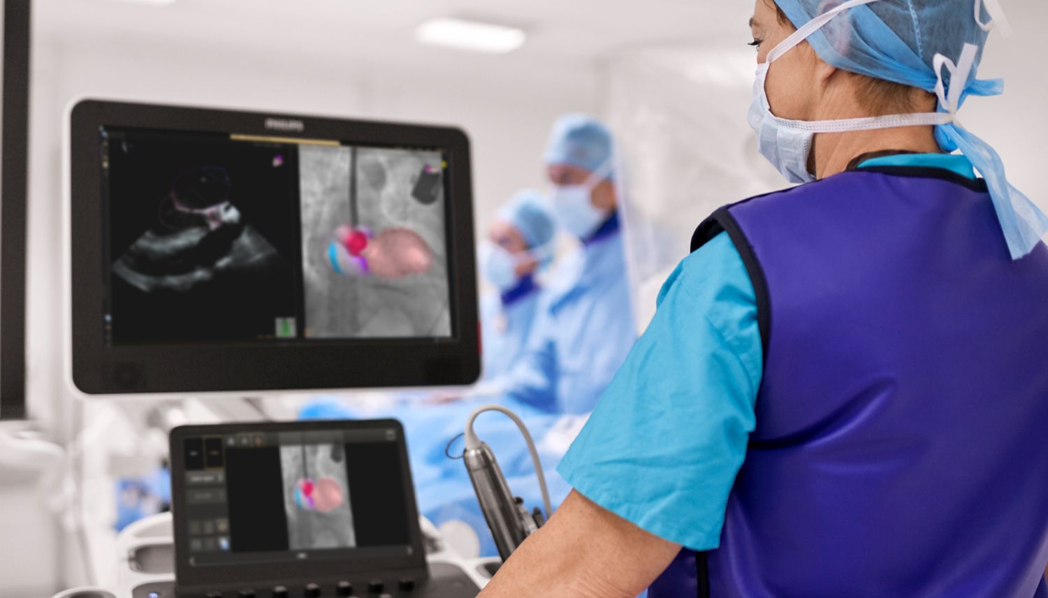
EchoNavigator significantly reduces transseptal puncture time[1].


Real-time fusion of echocardiography and fluoroscopyproved to be as safe and successful as standard best practice for TSP (transseptal puncture time). Moreover, efficacy was improved through significant reduction of time until TSP.1

10

years of innovations in fusion technology.

The Azurion system with the latest release of EchoNavigator, Release 4.0, builds on almost a decade of innovation in fusion technology and elevates your SHD procedures to the next level.

94%  

of clinicians.


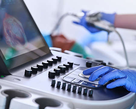
EPIQ MultiVue may help reduce the risk of choosing an incorrectly sized device2


 94% of clinicians who saw the new EPIQ CVxi thought the EPIQ MultiVue real-time alignment solution could help to reduce the risk of choosing an incorrectly sized device during interventional procedures2. 

17%  

reduction in procedure time can help you treat one more patient per day3


Treat one more patient per day by reducing procedure time by 17%3 with the optimized workflow options and clinical software of Azurion.

azurion c20 image

Azurion 7 M20 with FlexArm


Treat one more patient per day, by reducing procedure time by 17%3 with the optimized workflow options and clinical software of Azurion. 

Echo navigator image

EchoNavigator


Reorient and combine ultrasound and X-ray into a single augmented visualization. 

Echocardiography epiq image

EPIQ CVxi interventional ultrasound system


Premium interventional cardiology ultrasound system with powerful AI-based capabilities and nSight imaging to help you transcend today's complexities and propel interventional echocardiography into the next dimension. 

我們一直有興趣與您互動

告訴我們該怎麼幫助你

1
選擇您感興趣的領域
2
聯繫方式

Footnotes
 

*Reducing procedure time by 17%, with the ability to treat 1 more patient per day with optimized workflow options in image guided therapy and clinical software (Azurion - Philips Azurion Simulation Study 2016 - 12NC 452299123041 - FEB 2017). Results are specific to the institution where they were obtained and may not reflect the results achievable at other institutions. 

[1] Afzal, S., et al., First experience with real-time 3D anatomical fusion imaging during left atrial appendage occluder implantation. Eur Heart J Cardiovasc Imaging, 2017. 18(6): p. 719-720.This claim is based on a single center study with limited number of patients. This claim is limited to mitral clipping and left atrial appendage occlusion. 

[2] Jungen et al., PLoS One 2015 Oct 14;10(10); Left Atrial Appendage Closure Guided by Integrated Echocardiography and Fluoroscopy Imaging Reduces Radiation Exposure.

[3] Reducing procedure time by 17%, with the ability to treat 1 more patient per day with optimized workflow options in image guided therapy and clinical software (Azurion - Philips Azurion Simulation Study 2016 - 12NC 452299123041 - FEB 2017). Results are specific to the institution where they were obtained and may not reflect the results achievable at other institutions. 


Please note that not all products displayed on our website may be available in your country due to varying local regulations and market conditions. For the most accurate availability information, please contact your local representative. 

You are about to visit a Philips global content page

Continue

You are about to visit a Philips global content page

Continue

使用最新版 Microsoft Edge、Google Chrome 或 Firefox 瀏覽本網站效果最佳。