Bone Suppression image enhancement
Bone Suppression image enhancement technology
Features

Confident interpretation
By providing a soft tissue image in addition to a conventional chest image, Bone Suppression provides decision-making support without the need for additional X-ray dose or time. With Bone Suppression, actionable lung nodule detection is improved up to 16.8%. [1]

Efficient workflow
Bone Suppression does not require an additional procedure, add to examination time or require additional equipment. Depending on the protocol, for each adult erect chest PA/AP image a bone-suppressed image can be automatically generated and sent to PACS in addition to the conventional image. Both the conventional and the bone-suppressed image can be accessed and reviewed at the PACS viewing station at any time.
Bone Suppression image enhancement technology is available on
-
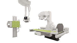
CombiDiagnost R90
This remote controlled fluoroscopy system in combination with high-end digital radiography is designed for consistent, superb image quality and high room utilization, in a cost effective manner.
709030
“Bone suppression technology helps us make the correct diagnosis more quickly and confidently than ever before.”
Jared Christensen, MD
Associate Professor of Radiology and Director of the Duke University Lung Screening Progam
Documentation
- Resources
-
Footnotes
*ClearRead Bone Suppression by Riverain Technologies [1] Freedman M et al. Improved detection of lung nodules with novel software that suppresses the rib and clavicle shadows on chest radiographs. Radiology. 2011. [2] See 15 more patients/day, save 8 hrs. overtime/week, and avoid approx. 28 retakes/week (compared to the previous release of DigitalDiagnost and based on 100 patients per day. Actual results in other cases may vary)


