FieldStrength MRI magazine
User experiences - March 2017
UVM appreciates latest neuro MR methods for diagnosing and workflow
The MRI staff at University of Vermont (UVM) Medical Center has evaluated recent methods to identify how their protocols for the brain can be improved. In these studies, they saw that these latest methods for susceptibility weighted imaging, motion reduction and perfusion imaging provide significant differences that benefit diagnostic confidence and are favorable for workflow. Their evaluation convinced them to incorporate SWIp, MultiVane XD and pCASL into their standard MR exams, which also helps them contribute to a first time right approach for their patients.
“We switched over entirely. SWIp is now included in all our routine brain exams”

Dr. Joshua Nickerson
Joshua Nickerson, MD, is the Medical Director of the University of Vermont MRI Research Center. His research interests include novel MRI-based perfusion techniques for functional brain imaging, high resolution MR imaging of the orbit, and new methods for using MRI to evaluate multiple sclerosis. He is also involved in several interdisciplinary studies in the UVM research community.

Dr. Richard Watts
Richard Watts, PhD, is Co-Director of the University of Vermont MRI Center for Biomedical Imaging, and an Associate Professor in the Department of Radiology. His academic interests include T1-weighted imaging in multiple sclerosis, Alzheimer’s disease, brain tumors, multimodal imaging of mild traumatic brain injury (mTBI), therapeutic hypothermia, and imaging connectomes using high b-value diffusion imaging.
Leading the way for neuro diagnostic imaging
As the UVM Medical Center in Burlington, Vermont, strives to provide a high level of patient care, the MRI team is always watchful for ways to increase diagnostic value. The team is driven to explore the added value of new methods, provided these help improve their exams and broaden the diagnostic scope of MRI. When additional methods for brain MRI became available to them, they systematically compared these methods with their previous standards to investigate the benefits and determine how these could help them expand their capabilities or improve their way of working.
SWIp in patients with hypertension, amyloid angiopathy, trauma
Neuroradiologist Joshua Nickerson, MD, discusses their findings in comparing SWIp versus T2* weighted imaging in different types of patients.
The SWIp sequence offers high resolution 3D susceptibility weighted brain imaging, which helps to visualize deoxygenated blood or calcium deposits. In combination with other clinical information, it may help in the diagnosis of various neurological pathologies
“With SWIp we are basically looking for blood byproducts. It is a sensitive method for visualizing small lesions containing deoxygenated blood. In our comparison, SWIp images are vastly better than gradient echo imaging, there’s no question of that anymore.” “We find the SWIp images very useful in three areas in particular. In patients with a history of hypertension, it offers clear visualization of hemosiderin deposition from hypertensive hemorrhages. We certainly see a greater number of foci of hemosiderin deposition on the SWIp images than on the T2* gradient echo images. In addition, it also helps us visualize amyloid depositions in patients with amyloid angiopathy.” Dr. Nickerson mentions trauma patients are the third large area where SWIp is useful. “We benefit from SWIp in trauma patients, certainly in cases with diffuse axonal injury and shearing injuries. Our study shows that SWIp usually provides us better visualization,” he says. “Apart from these three, SWIp also helps us to beautifully depict the normal venous anatomy in patients with venous outflow issues or vascular congestion. In some cases, we have seen downstream effects of arterial problems. And in patients with vascular malformations we have seen deposition of blood products associated with those.”
“Our study shows that SWIp usually provides better visualization than gradient echo imaging in trauma patients”
SWIp in all brain exam protocols at UVM
“We switched over entirely. SWIp is now included in all our routine brain exams. We developed two different SWIp sequences: a high spatial resolution (0.5 x 0.5 mm) version that takes 5.5 minutes and our fast SWIp that takes just three minutes. Only in patients that are moving tremendously we occasionally still acquire a gradient echo sequence.” “For us, SWIp use has resulted in more diagnostic confidence when small lesions, such as small shear injuries, vascular malformations, or minute amounts of calcification, need to be detected,” says Dr. Nickerson. “Our physicians greatly value the SWIp images. When we get patients transferred from other facilities with SWIp missing from their exam, we have several neurologists and neurosurgeons who order a new MRI exam because they want to see the SWIp images.”
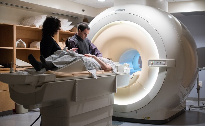

University of Vermont MRI Center
“For us, SWIp use has resulted in more diagnostic confidence when small lesions need to be detected”
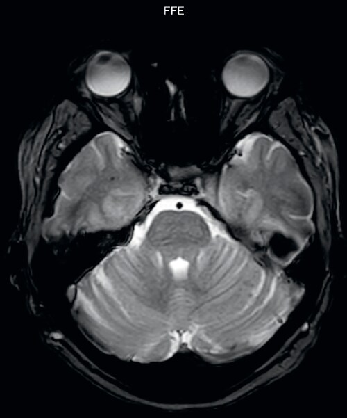
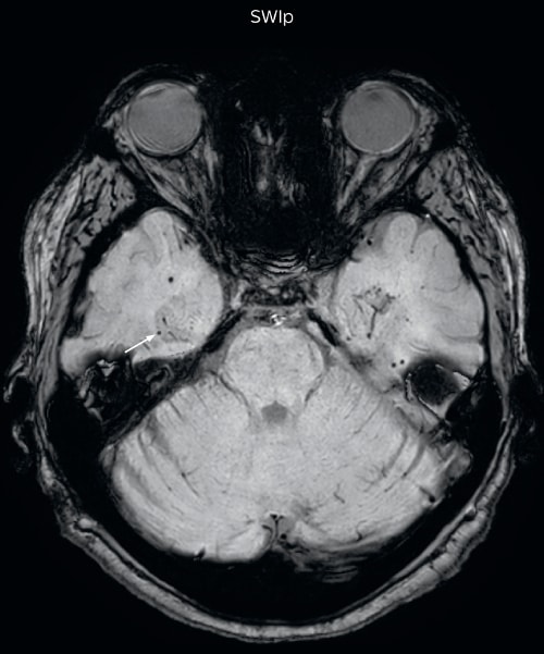
Hemosiderin foci in brain
Gradient echo imaging and SWIp are compared in a patient with radiation-induced foci of hemosiderin deposition. A greater number of small foci is seen on the SWIp image. Ingenia 3.0T
“MultiVane XD is especially useful for imaging patients with diseases that cause white matter changes on T2-weighted images”
Diagnostic imaging in presence of motion
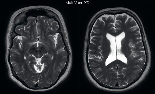
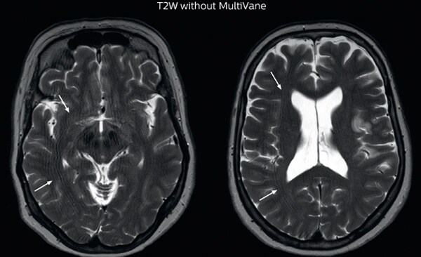
“Motion artifacts can obscure subtle findings, make image interpretation more difficult and decrease diagnosis confidence. For example, when imaging the cerebellum or brain stem, or when looking for subtle multiple sclerosis (MS) lesions, motion can be problematic,” says Dr Nickerson. MultiVane XD motion-free imaging delivers diagnostic images even in the case of severe patient motion. A more relevant patient group is one with typical small artifacts related to moderate motion like an occasional cough. The absence of those artefacts brings forth better day-to-day diagnostic confidence. MultiVane XD works in multiple orientations and for various contrasts, such as T1-weighted, T2 weighted and FLAIR. Trevor Andrews, PhD, explains that the team compared motion artifacts seen in the brain with MultiVane XD and with T2-weighted TSE. “In nine out of the ten datasets in our studywe saw clear improvementswith MultiVane XD, while in the tenth dataset image qualitywas comparable. The MultiVane XD sequence is now used in the majority of patients that present at UVM for brain MRI.”
“Motion artifacts can obscure subtle findings”
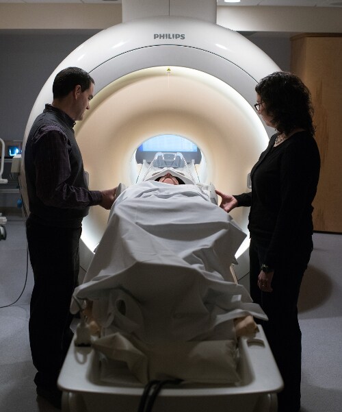
Motion-free imaging of white matter changes with MultiVane XD
“We saw MultiVane XD provide remarkable improvement, not only for artifacts caused by patient motion, but also for the extent of pulsation artifacts in the basal cisterns. Based on these results, we have added the MultiVane XD sequence to our brain studies,” says Dr. Nickerson. “MultiVane XD is especially useful when imaging patients with diseases that cause white matter changes on T2-weighted images, such as MS, small vessel disease, vasculitis and sarcoidosis,” says Dr. Nickerson. “Many of these are only visible on T2-weighted or FLAIR images, and sometimes aren’t even seen with FLAIR images. However, when using MultiVane XD and we don’t see any motion on the rest of the scan, but still do see a signal abnormality, we can probably attribute that to a real disease process, rather than an artifact.”
If patient motion would not hamper workflow
As workflow can suffer significantly from patient motion, a good motion reduction technique is also desirable from that perspective. According to UVM technologist Sarah Comtois, technologists used to spend much time on communicating with patients who tended to move, supplying extra padding for the head, re-running sequences, and consulting with radiologists to ensure that the results were acceptable. For Dr. Nickerson, MultiVane XD adds substantial value: “It’s not just time being saved on the technologist’s side,” he says. “If the use of MultiVane XD results can help in reducing calls back to the reading room to evaluate images of questionable diagnostic value, there are significant time savings there, too.” Dr. Nickerson suggests that MultiVane XD may have the potential to help them shorten time slots for pediatric patients. “That population is particularly prone to motion, so sometimes larger gaps are planned between slots with the understanding that sequences may have to be repeated and the study may take longer than it would in an adult. There may be a significant impact on time saving if we didn't have to take that into consideration''
“It’s not just time being saved on the technologist’s side”
MRI motion artifact reduction in brain
The images made with MultiVane XD show significant reduction in motion artifact compared to the T2-weighted images without MultiVane below them. Scanned on Ingenia 3.0T
“In the cases in our study, pCASL was at least as representative as perfusion with contrast agent”
Fast brain perfusion imaging without contrast agent
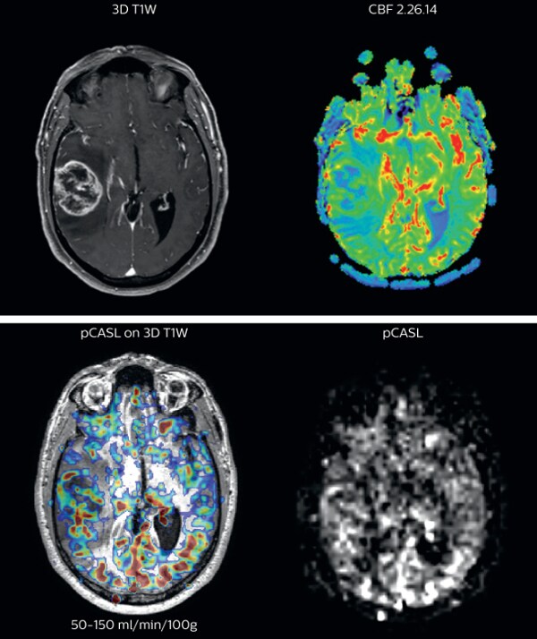
Brain perfusion imaging is typically performed using a contrast agent. However, pCASL allows visualization of brain perfusion and physiology without contrast agent injection. This fast sequence can be an alternative for perfusion imaging in patients who are contraindicated for use of gadolinium based contrast agents. “We have compared pCASL to T2*-weighted perfusion imaging with contrast agent in patientswith brain tumors”, says Dr. Nickerson. “In the cases included in our study, pCASL was at least as representative as perfusion with contrast agent. It’s a pretty big improvement if we’re avoiding giving gadolinium and we’re getting a quite equivalent dataset, that’s a pretty big improvement.”
“If a patient moves during pCASL perfusion imaging, we can simply repeat the scan because there’s no contrast injection involved”
pCASL broadly used in brain tumor patients
The pCASL method is now broadly used at UVM. “It’s a short sequence, and is ideal for use in patients where motion is a concern. pCASL is currently included in all our MRI exams for patients with a known tumor, either initial or post-operative and all follow-ups. Additionally, it can also be used when we examine for stroke. And of course, pCASL is an alternative allowing perfusion imaging in patients with compromised renal systems, for whom contrast agent is contraindicated.” “Furthermore, we image a fair number of pediatric tumors here and the repeatability of pCASL is a great benefit when scanning pediatric patients with brain tumors. If the patient moves during the acquisition of a DSC perfusion scan, we missed our shot. However, if a patient moves during pCASL, we can simply repeat the scan because there’s no contrast injection involved,” says Dr. Nickerson Richard Watts, PhD, brings forward another aspect. “Quantification with pCASL does not have the issue of selecting an arterial input function like with contrast-agent based scans. In the brain tumor studies that we’ve been running, we feel it’s not sufficient just to ascertain one side has got more blood flow than the other side. In the future, we want to move towards quantitative comparisons between subjects.”
Cerebral blood flow in glioblastoma
The pCASL perfusion map overlaid on the 3D T1 image demonstrates a peripheral rim of elevated cerebral blood flow corresponding to the centrally necrotic glioblastoma. The pCASL-generated CBF closely approximates the rim of elevated rCBV obtained with DSC contrast-enhanced perfusion imaging. Scanned on Achieva 3.0T dStream
A boost in diagnostic confidence
The MRI staff at UVM feels that the adopted methods not only boost their diagnostic confidence, but also contribute to improving workflow. Two of the methods, SWIp and MultiVane XD, are now used in virtually all brain MRI scans performed at the institute. Additionally, pCASL is considered a robust addition for brain perfusion imaging, providing an option for fast and easy repeat scanning if patient movement occurs, and offering an alternative for patients who do not tolerate contrast agent use. “Our experience is that these methods help us in obtaining the best possible scans for our patients. I would recommend to anyone to use these methods if they have them at their disposal,” says Dr. Nickerson.
“I would recommend to anyone to use these methods if they have them at their disposal”
Results from case studies are not predictive of results in other cases. Results in mother cases may vary.
More from FieldStrength
