I want to learn more
Stay up-to-date and get informed about interesting topics. Or get in touch with our sales department.

Addressing the obstacles to confident diagnosis:
Inconclusive studies
Too many imaging studies still result in inconclusive reports due to poor visualization or nonvisualization of anatomical structures, which can delay diagnosis and treatment, add stress for patients and staff and increase costs. The usual suspects for compromised image quality are well known: acquisition parameter errors, motion, fat and metal artifacts, operator error, and technology limitations.
The good news: we are making significant progress against the most challenging obstacles to diagnostic confidence in every imaging modality. Here are some examples of what that looks like.
What you'll see
")
Fine-tuning fluoroscopy Learn more about Dynamic UNIQUE:
")
Delivering diagnostic certainty in CT Learn more about IQon Spectral CT:
")
Revealing more with digital PET Learn more about Vereos digital PET/CT:
")
New perspectives in MRI Learn more about Multivane XD:
Fine-tuning fluoroscopy
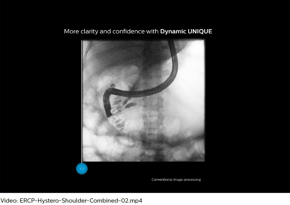
Addressing quantum noise
Modern fluoroscopy poses significant challenges for digital image processing. Low-dose settings are commonly used today in clinical practice, particularly for children, who are most sensitive to the damaging effects of ionizing radiation. While a reduction in dose is always desirable, these settings inevitably result in images with high quantum noise, which may be difficult for radiologists to read and can result in longer fluoroscopy times.
Dynamic UNIQUE combines noise suppression by temporal averaging (inter-frame) and spatial noise suppression (intra-frame). Temporal averaging, which accumulates the dose of successive frames, results in a depiction of static anatomical structures that mimics high-dose acquisition. However, it may fail in areas of motion. Dynamic UNIQUE suppresses temporal averaging when movement is detected preventing lag and shadowing of moving structures.
In moving regions of the image and if the sequences are very short, Dynamic UNIQUE applies spatial noise suppression. It uses a physical noise model to assess the contrast-to-noise ratio, which allows for structure-adaptive noise suppression within a single frame. The strength of the spatial noise suppression is adapted to the strength of the temporal noise reduction in adjacent areas. As a result, the image noise level is kept consistently low over the entire image.
Reduced noise impression results in improved detail visibility. Anatomical details are preserved in every image, with no artifacts of moving structures, no lag effect and no shadows of catheters, tubes and lines. Radiologists experience less risk to miss important details, leading to more confidence in the diagnosis.
")
Addressing quantum noise
Modern fluoroscopy poses significant challenges for digital image processing. Low-dose settings are commonly used today in clinical practice, particularly for children, who are most sensitive to the damaging effects of ionizing radiation. While a reduction in dose is always desirable, these settings inevitably result in images with high quantum noise, which may be difficult for radiologists to read and can result in longer fluoroscopy times.
Dynamic UNIQUE combines noise suppression by temporal averaging (inter-frame) and spatial noise suppression (intra-frame). Temporal averaging, which accumulates the dose of successive frames, results in a depiction of static anatomical structures that mimics high-dose acquisition. However, it may fail in areas of motion. Dynamic UNIQUE suppresses temporal averaging when movement is detected preventing lag and shadowing of moving structures.
In moving regions of the image and if the sequences are very short, Dynamic UNIQUE applies spatial noise suppression. It uses a physical noise model to assess the contrast-to-noise ratio, which allows for structure-adaptive noise suppression within a single frame. The strength of the spatial noise suppression is adapted to the strength of the temporal noise reduction in adjacent areas. As a result, the image noise level is kept consistently low over the entire image.
Reduced noise impression results in improved detail visibility. Anatomical details are preserved in every image, with no artifacts of moving structures, no lag effect and no shadows of catheters, tubes and lines. Radiologists experience less risk to miss important details, leading to more confidence in the diagnosis.
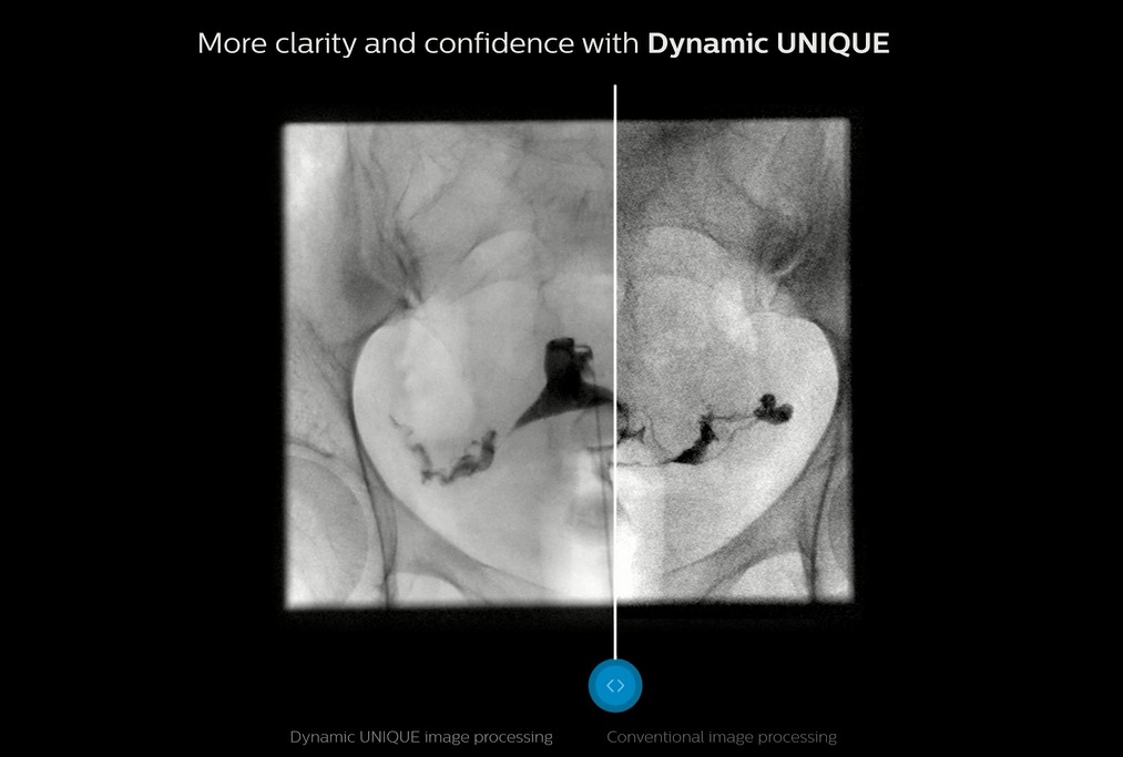
Managing contrast and brightness impression
Because fluoroscopy means live viewing, relevant diagnostic information in the images must be visible straightaway at the correct level of brightness and contrast. Furthermore, because the human eye is very sensitive to temporal variations, the contrast and brightness impression must be stable over time. This is particularly challenging in view of the rapid temporal variations occurring in fluoroscopy, particularly with respect to:
- The body region under examination, e.g. when examining the esophagus
- The field-of-view, e.g. when moving the patient or modifying the collimated area
- Areas of direct radiation
- The distribution of the contrast agent
- The dose level
When the dose level or the field-of-view is modified (e.g. by modifying its size, by shifting it from the body center to the periphery, or by moving the patient), Dynamic UNIQUE automatically determines the signal level in the anatomical region and maps it to an appropriate brightness level. Areas of direct radiation are reliably detected and excluded from the brightness adjustment. In contrast, simpler methods include areas of direct radiation in adjustments, resulting in dark images and obscured anatomy, as well as annoying flickering.
Optimal image stability for fluoroscopy and spot images at any time leads to comfortable viewing of high-quality images, less time needed to adapt to the luminosity, and ultimately, a smooth and efficient workflow.
")
Delivering diagnostic certainty in CT
Tackling indeterminate scans
Conventional CT scans often produce ambiguous or inaccurate data resulting in a need for additional testing. For example, in cases where a conventional image from a scan lacks the clarity typically provided by IV contrast, the resulting scan can be indeterminate, necessitating additional exams and resources.
IQon Spectral CT is the world’s first and only detector-based spectral CT which consistently captures spectral information without special planning or set-up.
Diagnostic certainty exists when multiple layers of spectral data allow the first exam to be the right exam, whether it’s with MonoE images, iodine images, virtual non-contrast images or Z effective images. In addition, IQon Spectral CT has been shown to reduce average scan cost.*
* Norwood, Don, MD, MBA. Economic impact of IQon for patients with renal insufficiency. 2017. Data on file.
")
Image courtesy of CARTI Little Rock, Arkansas, USA
Clinical case: A patient arrives in the ER with abdominal pain and is scanned to determine the cause. The conventional image result was indeterminate (left); however, because the patient was scanned on the IQon (right) using the spectral Low Mono E image the contrast visualization was improved.
")
Images courtesy University Hospitals, Cleveland, Ohio, USA
Identifying pulmonary emboli
Small pulmonary emboli (PE) can be difficult to identify on conventional CT images alone, and perfusion defects in the area of the PE can be overlooked.
The IQon Spectral CT aids the radiologist by providing spectral results that can help identify perfusion defects. Multiple spectral results available with every patient scanned on the IQon Spectral CT can aid the radiologist in the ability to identify multiple perfusion results.
In this example, the patient presented with a suspected PE. After reviewing the conventional scan, spectral results were applied, resulting in multiple perfusion defects being identified by the radiologist in both lungs.
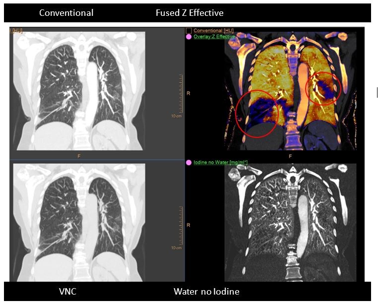
Images courtesy University Hospitals, Cleveland, Ohio, USA
Revealing more with digital PET
Faster scans, less discomfort
While PET/CT is the modality of choice for large-anatomical-area lesion detection, acquisition time for analog systems (15-20 minutes) can result in patient discomfort and associated motion artifacts that compromise image quality.
Vereos Digital PET/CT is the world's first and only true digital PET/CT system, which positions radiologists to go beyond current limitations to improve patient care. Vereos’ Digital Photon Counting technology enables uncompromised lesion detectability at 1/10th the time.* With this technology, radiologists can perform a whole body FDG study in under 5 minutes.
Given Vereos’ increased sensitivity and lesion detection in less time, radiologists may adjust acquisition times depending on their need to accommodate patient comfort.
* Zhang, J, Evaluation of speed of PET acquisition: How fast can we go? - A validation of list mode PET simulation approach with true acquisitions, SNMMI 2017.
")
Equivalent detectability of neck lesion at 9 sec/bed, relative to a 90 sec/bed acquisition.
Sample images acquired in a clinical study of the Vereos PET/CT system at The Ohio State University. Investigational device limited by law to investigational use.
New perspectives in MRI
Addressing breathing motion artifacts
Imaging of the shoulder is susceptible to breathing motion artifacts, particularly when scanning in axial orientation.
Using MultiVane XD, Philips makes it possible for the radiology staff to deliver imaging with motion reduction through robust motion correction. Its extended reconstruction algorithm analyzes and corrects blades so that image distortions due to both translational and rotational components of rigid body motion are removed. Additionally, the k-space center of blades corrupted with through-plane motion is removed to further decrease motion artifacts.
MultiVane XD makes it possible for radiologists to stick to their basic sequence optimization steps for obtaining high-quality images. By providing motion correction in short scan times, MultiVane XD delivers high resolution diagnostic images even in the case of severe patient motion.
")
Comparison of axial fat suppressed PD-weighted imaging with (left) and without (right) MultiVane XD in the left shoulder demonstrates imaging with MultiVane XD provides excellent image quality, even in presence of motion.
")
510K pending. Not available for sale in the USA.
By using Philips’ operationally intuitive solution, radiologists can experience up to 50% reduction in exam time for anatomic contrasts in all anatomies, with virtually equal image quality.
Reduce exam time without compromising image quality
Shortening MR exams without compromising image quality is a pressing challenge in radiology today. Whether in acute care or non-urgent diagnostic circumstances – and for reasons ranging from patient stress to operational costs and throughput – shorter is better. How do you reduce exam time without compromising image quality?
With our innovative MR solution Compressed SENSE, radiology departments can acquire all 2D and 3D scans faster without compromising imaging quality. Philips Compressed SENSE is a breakthrough acceleration technique that speeds up not only sequences, but also the entire exam.
Learn how Philips solutions help radiologists overcome obstacles to confident diagnosis. See them all: