Multi Modality Viewer: single platform for all your viewing needs
IntelliSpace Portal 10 covers a host of your clinical needs going beyond neurology, cardiology, and oncology to include a range of additional domains.
- 3D Modeling
-
3D Modeling
Streamlined modeling workflow
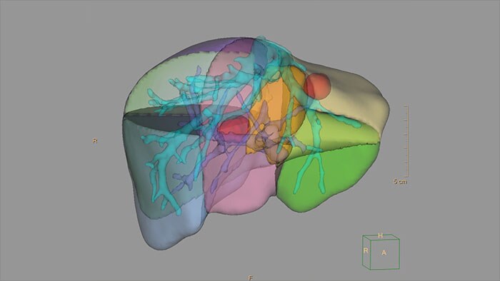
Allows to view volumetric images of anatomical structures, perform segmentation, edit and combine segmented elements (tissues) into a 3D model.
Benefits
- Studies of CT & MR can be used for creating a single 3D model of the same patient. The application provides tools that allow the user to align between the volumes of interest in the images.
- 3D Modeling batches files can be easily exported in standard formats such as STL, with the option to also provide a 3D PDF as an additional means for results sharing with 3D printing or other services* .
- The user may determine the information related to the exported elements of the 3D model such as smoothness and output mesh size.
- Contours can also be exported as RT Structures.
*3D models are not intended for diagnostic use.
- Advanced Vessel Analysis (AVA)
-
Multi Modality Advanced Vessel Analysis (AVA)
Comprehensive vascular analysis planning
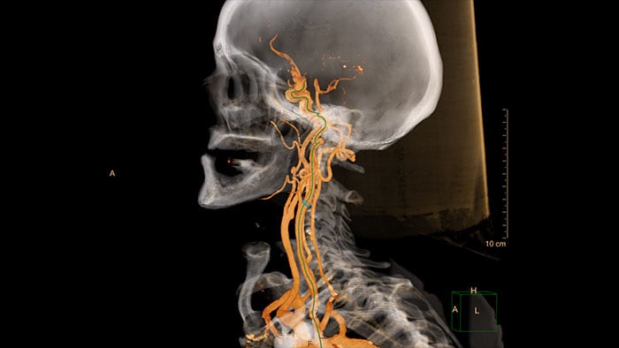
Designed to examine and quantify different types of vascular lesions from CTA and MRA scans. It accommodates different modes of inspection, allows labeling different vascular lesions, and helps navigating through multiple findings.
Demonstrated to reduce the post-processing time by 50% when compared to manual Head & Neck CT angiography (CTA) analysis*.
Benefits
- Ability to choose which Head & Neck Bone Removal method to be used (Standard vs. Smooth).
- Customizable Volume rendering “smoothness” for the 3D Head & Neck vascular structure using a smoothness control.
* Ardley N et al. Efficacy of a new post processing workflow for CTA head and neck. ECR 2013 / C-1760.
- Tumor Tracking
-
Multi Modality Tumor Tracking (MMTT)
Streamlined workflow for follow up and analysis of oncology patients
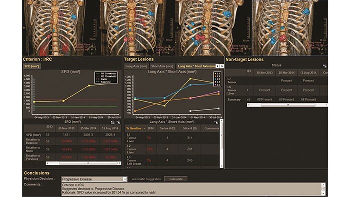
MMTT is a post processing software used to display, process, analyze and quantify anatomical and functional images, for CT, MR, PET/CT, SPECT/CT and Dual Energy CT at one or multiple time points.
Benefits
- Enhanced semi-automatic volumetric segmentation.
- Selectable oncology response criteria including standards such as RECIST 1.0, RECIST 1.1, WHO, CHOI, PERCIST, irRC and mRECIST, as well as PET SUV analysis including glucose-corrected SUV.
- Findings can be shared with other IntelliSpace Portal applications such as CT Liver Analysis and CT Viewer or exported in different formats including RT Structures.
- Tumor Tracking qEASL
-
Multi Modality Tumor Tracking qEASL (MMTT qEASL)
Semi-automatic tumor quantification
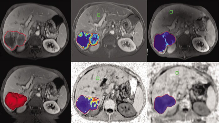
This semi-automated 3D (Volumetric) tumor response assessment tool, based on EASL (European Association for the Study of the Liver) criteria incorporates functional information from contrast-enhanced scans.
Benefits
- Multi Modality Tumor Tracking supports the creation of Quantitative EASL (qEASL) maps used to measure segmented volumes of interest (VOI) in heterogeneous lesions.
- Data are presented as color map overlaid on the scans to show regional tumor enhancement heterogeneity. The color regions of the segmented lesions are where there is more enhancement than the pre-defined reference region.
- Viewer (MMV)
-
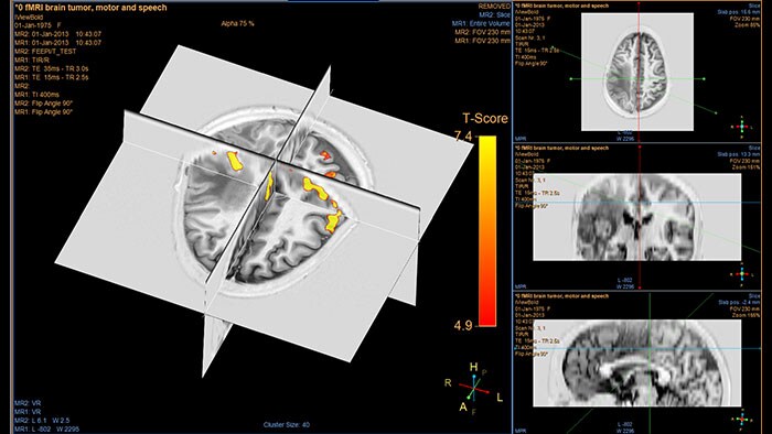
Multi Modality Viewer (MMV)
Initial viewing platform for advanced analysis needs
Supports study review, side-by-side comparison, series arrangement as well as 2D and 3D manipulation of MR, CT, PET, NM, US, DX, CR, RF and XA images.
Benefits
- Supports multiple image rendering modes and geometries as well as fusion capabilities of two series including registration options.
- Offers a set of tools for basic measurements, stitching multi-station data and generation of new DICOM series/objects for communication purposes.
- Supports the generation of MR DICOM series in the form of a dedicated MPR series derived from the 3D T1 acquisition, fused with objects like fiber, SPM (fMRI) and/or segmented structure.
- A unique patient-centric workflow facilitates communication between the IntelliSpace Portal and Philips Image Guided-Therapy systems, to automatically launch relevant advanced analysis data before intervention*.
* This requires specific plug-in installation on the ISP client which integrates with the Philips Cath-lab systems.
- Zero Foot Print Viewer
-
Zero Footprint Viewer*
Access to advanced DICOM viewing anywhere
Provides a clinically rich viewing environment, such as quick prior comparison with automatic registration, MPR and Volume modes and Key images workflow. The HTML based viewer allows access anywhere to imaging data stored and created on IntelliSpace Portal outside and inside the hospital.

Benefits
- Built in Peer2Peer. Real-time collaboration capabilities supports communication and consultation between physicians.
* The viewer is not intended for diagnostics image review. Viewer is supported on OS X 10.10 and Windows 7,10 using: Internet Explorer, Chrome, Edge, Safari. Functionality is not available in IX workstation configuration.
